

现期刊物2026
卷册: 16, 期号: 13
生物化学
An Immunoprecipitation-Based Nonradioactive Kinase Assay to Measure Akt Kinase Activity in Mammalian Cell Lines
基于免疫沉淀法检测哺乳动物细胞系中Akt激酶活性的非放射性激酶实验
NADH-Dependent Oxidoreductase Activity Assay of OsAIM1 Using a Microplate Reader
利用酶标仪测定 OsAIM1 的 NADH 依赖性氧化还原酶活性
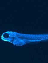
An Optimized Protocol for the Characterization of Zebrafish ApoB-Containing Lipoproteins Using the LipoGlo System
利用 LipoGlo 系统表征斑马鱼含 ApoB 脂蛋白的优化实验方案
生物信息学与计算生物学
Multiply Perturbed Response: A Computational Protocol to Identify Cooperative Allosteric Residue Combinations Driving Protein Conformational Transitions
多重扰动响应法:识别驱动蛋白质构象转变的协同变构残基组合的计算方案
Simultaneous Transcriptomic Analysis of Both Host and Symbiont in Insect–Fungus Interactions
昆虫与真菌相互作用中宿主和共生体的同步转录组分析


Efficiency-Corrected Relative Quantification of qPCR Data Using LinRegPCR and a Spreadsheet-Based Workflow
利用 LinRegPCR 和电子表格工作流程对 qPCR 数据进行效率校正的相对定量
癌症生物学
Construction and Functional Evaluation of Cyclic Peptide-Based CAR T Cells in Tumor Models
基于环肽的 CAR-T 细胞构建及其在肿瘤模型中的功能评价
Generation and Characterization of Adaptive Anoikis-Resistant Cells Using Cyclic Attachment-Detachment Culture of Cancer Cells
利用癌细胞周期性贴壁脱附培养构建并表征适应性抗失巢凋亡细胞
细胞生物学
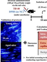
Quantitative Assessment of Capillary Permeability in Deep Intracardiac Capillaries Using Fluorescent Dextran
利用荧光葡聚糖定量评估心脏深部毛细血管的通透性
发育生物学
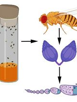
Visualizing Membrane Nanotube Dynamics in Drosophila Oocyte Using Live-Cell Imaging
利用活细胞成像观察果蝇卵母细胞中膜纳米管的动态变化
环境生物学
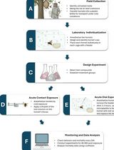
Acute Contact and Oral Testing of Chemical Compounds on Vespa velutina nigrithorax (Hymenoptera, Vespidae) Under Laboratory Conditions
实验室条件下化合物对墨胸胡蜂的急性接触和经口毒性试验

免疫学
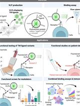
A Flow Cytometry–Based Assay to Quantify the Binding of Transmembrane Ligands to Their Cognate Receptors Using Fluorescent Virus-Like Particles
一种基于流式细胞术利用荧光病毒样颗粒定量检测跨膜配体与其相应受体结合的方法
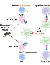
Improved Protocol for Establishing CD4+ Hybridomas Specific for Human Class II MHC/Peptide Complex
建立人Ⅱ类 MHC 肽复合物特异性 CD4+ 杂交瘤的改进方案
分子生物学
CRISPR-PITA: An Imaging-Based CRISPR/dCas9 Assay to Determine Recruitment Directionality of Nuclear Proteins
CRISPR-PITA:一种用于确定核蛋白招募方向性的基于成像的 CRISPR/dCas9 检测方法
神经科学
Whole-Mount Immunostaining of Tyrosine Hydroxylase for Dopaminergic Neuron Analysis in Zebrafish Larvae
斑马鱼幼体酪氨酸羟化酶整体免疫染色,用于多巴胺能神经元分析
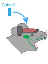
Hemispherectomy-Based Optical Window for In Vivo Visualization of Trigeminal Ganglion Neurons in Mice
基于大脑半球切除术建立光学窗口,实现小鼠三叉神经节神经元的活体成像
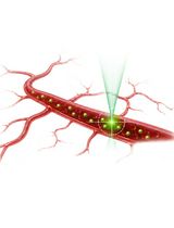
Protocol for In Vivo Two-Photon FCS to Measure Nanocarrier Number and Flow Velocity in Mouse Cerebral Microvasculature
利用活体双光子荧光相关光谱测量小鼠脑微血管中纳米载体数量与流动速度的实验方案

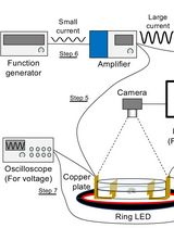
Tracking AC Electric Stimulation–Induced Persistent Locomotion Behavior in the Nematode Caenorhabditis elegans
追踪交流电刺激诱导的秀丽隐杆线虫持续性运动行为
植物科学
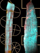
4D Imaging of Brown Algal Cells
褐藻细胞的四维成像

A Dual-gRNA CRISPR/Cas9 System for Efficient Generation of Large Fragment Deletions in Poplar
一种用于在杨树中高效构建大片段缺失的双 gRNA CRISPR/Cas9 系统
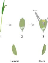
Lodicule Isolation and Morphometric Analysis During Rice Floret Opening
水稻小花开放过程中浆片的分离与形态测量分析
干细胞
Stepwise Differentiation of Mouse Embryonic Stem Cells Into Murine Blood Vessel Organoids With Endothelial Lineage Tracing for Quality Control
将小鼠胚胎干细胞逐步分化为小鼠血管类器官并通过内皮细胞谱系追踪进行质量控制