往期刊物2026
卷册: 16, 期号: 9
生物化学
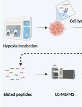
A Suspension-Trapping Protocol for Bottom-Up Proteomics Sample Preparation
基于悬浮捕获法的自下而上蛋白质组学样品制备方案
Reconstitution of Active Plant H+-ATPase AHA2 in Giant Unilamellar Vesicles
在巨大单层囊泡中重构具有活性的植物 H⁺-ATPase AHA2
生物物理学
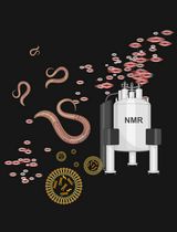
Lipid Analysis in Live Caenorhabditis elegans Using Solution-State NMR Spectroscopy
利用溶液态 NMR 光谱分析活体秀丽隐杆线虫中的脂质
细胞生物学
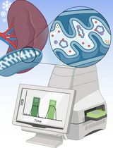
Assessing Mitochondrial Respiratory Complex-Associated Function From Previously Frozen Mouse Placental Tissue
利用冻存小鼠胎盘组织评估线粒体呼吸复合体相关功能
A Cell-Based Protocol to Assess Manganese Content and Relative Transport Activity of Manganese Transporters
基于细胞体系评估锰含量及锰转运蛋白相对转运活性的实验方案
分子生物学
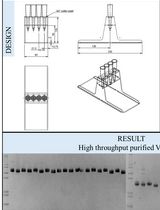
Parallelised Cloning, Mammalian Cell Expression, and Purification of Nanobodies Identified by Phage Display
噬菌体展示筛选纳米抗体的并行化克隆、哺乳动物细胞表达与纯化
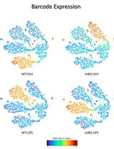
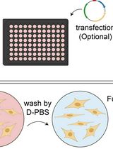
An Advanced Single-Cell RNA Sequencing (scRNA-seq) Protocol Utilizing Custom-Designed Multiplexing
基于自定义多重标记策略的高级单细胞 RNA 测序(scRNA-seq)方案
神经科学
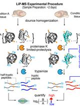
Limited Proteolysis Mass Spectrometry to Identify Protein Structural Differences in Brain Tissue
利用有限蛋白水解质谱鉴定脑组织中的蛋白质结构差异
植物科学
Kinetic Determination of Cytochrome b6f Activity In Vitro
体外动力学测定细胞色素 b6f 复合体活性
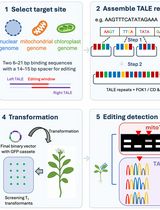
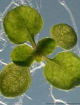
TALENs and Related Technologies for Editing Nuclear and Organellar Genomes in a Model Plant, Arabidopsis thaliana
TALENs及相关技术在模式植物拟南芥核基因组与细胞器基因组编辑中的应用
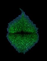
Detecting Touch-Induced Calcium Dynamics With Live-Cell Imaging in Torenia Stigma
利用活细胞成像检测蓝猪耳柱头中的触碰诱导钙动态变化
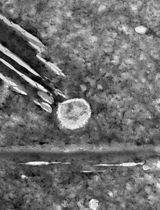
Extraction and Isolation of Extracellular Vesicles From Piper betle Leaves Using the Apoplastic Fluid Washing and Size Exclusion Chromatography Method
基于质外体液洗脱与尺寸排阻色谱法提取和分离蒌叶来源的细胞外囊泡

Quantitative Assessment of Heat Shock-Induced Ferroptosis-Like Cell Death via Electrolyte Leakage in Arabidopsis thaliana Seedlings
拟南芥幼苗热激诱导铁死亡样细胞死亡的电解质渗漏定量评估
干细胞
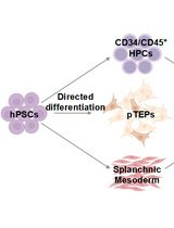
Generation of Functional Patient-Specific Thymus Organoids From Human Pluripotent Stem Cells (hPSCs) Using Air–Liquid Interface Culture
利用气液界面培养从人多能干细胞(hPSCs)构建患者特异性功能性胸腺类器官