现期刊物2026

卷册: 16, 期号: 13

left

Jul

5

Jun

20

Jun

5

May

20

May

5

Apr

20

Apr

5

Mar

20

Mar

5

Feb

20

Feb

5

Jan

20

Jan

5

right

生物化学

基于免疫沉淀法检测哺乳动物细胞系中Akt激酶活性的非放射性激酶实验

An Immunoprecipitation-Based Nonradioactive Kinase Assay to Measure Akt Kinase Activity in Mammalian Cell Lines

基于免疫沉淀法检测哺乳动物细胞系中Akt激酶活性的非放射性激酶实验

AP Amber Peek
JM Jay N. Mehta
DB Deepali Bhandari
51 Views
Jul 5, 2026
Protein kinase B, more commonly known as Akt, is a family of three serine/threonine kinases (Akt1, Akt2, and Akt3) that play a central role in regulating processes such as proliferation, survival, metabolism, and migration through phosphorylation of downstream targets. Given its involvement in numerous cellular processes, aberrant Akt signaling is prevalent across multiple cancer types, underscoring the need for Akt kinase assays to assess activity, regulatory mechanisms, and the efficacy of targeted interventions. Most existing Akt kinase assays rely on expensive commercial kits, some of which employ pre-purified, constitutively active Akt expressed in insect cells, bypassing physiologic autoinhibition of Akt; therefore, they are not suitable for evaluating allosteric inhibitors or context-dependent regulation. Here, we describe a detailed, step-by-step protocol for a nonradioactive Akt kinase assay using epitope-tagged, recombinant Akt1 expressed in a mammalian cell line and isolated by immunoprecipitation. This method eliminates the need to co-express Akt with upstream regulatory kinases or to purify active enzyme from insect cells, a time-consuming and technically demanding process, particularly when analyzing multiple Akt mutants. Because Akt is assayed in a regulated, autoinhibited state, this protocol enables direct evaluation of allosteric inhibitors that cannot be assessed using active Akt purified from insect cells. We note, however, that Akt1 kinase activity in this assay is measured from epitope-tagged, transiently overexpressed protein, which could influence cellular signaling dynamics. Despite this limitation, the cellular context preserves key regulatory features of Akt1 autoinhibition and membrane-dependent activation that are absent in assays using purified, pre-activated kinase. Together, this protocol supports analysis of Akt kinase activity under diverse experimental conditions, including receptor stimulation, pharmacologic treatment, allosteric inhibitor exposure, and mutations, using an accessible, economical, and physiologically relevant approach.
利用 LipoGlo 系统表征斑马鱼含 ApoB 脂蛋白的优化实验方案

An Optimized Protocol for the Characterization of Zebrafish ApoB-Containing Lipoproteins Using the LipoGlo System

利用 LipoGlo 系统表征斑马鱼含 ApoB 脂蛋白的优化实验方案

MH Monica R. Hensley
SF Steven A. Farber
42 Views
Jul 5, 2026
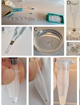
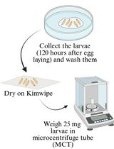
Apolipoprotein B–containing lipoproteins (ApoB-LPs) transport lipids throughout the circulation and are closely associated with cardiovascular disease in humans. Many aspects of ApoB-LP biology remain elusive, often due to their indirect characterization through the measurement of plasma triglycerides and cholesterol. The conventional approach provides limited information on ApoB-LPs number and size distribution, essential features that influence cardiovascular disease risk. Additionally, drug studies have historically been limited to the use of mammalian research models, which are not suited for high-throughput experiments. Therefore, we generated a reporter system (LipoGlo) utilizing a luciferase enzyme (NanoLuc) fused to the C-terminus of the zebrafish (Danio rerio) ApoBb.1 protein. In metazoans, ranging from insects to humans, each ApoB-LP contains a single ApoB molecule, such that the luminescence emitted from these transgenic fish is proportional to the total number of ApoB-LPs. The LipoGlo zebrafish reporter generates a quantitative chemiluminescent signal that can be used in plate-based assays to measure lipoprotein quantities, a gel-based assay that can measure lipoprotein size distribution, and chemiluminescent microscopy that can, for the first time, visualize lipoprotein localization in a larval zebrafish. LipoGlo, combined with the amenability of zebrafish to genetic approaches, facilitates the rapid assessment of any gene or drug’s role in ApoB-LP molecular and cell biology. This protocol describes three optimized LipoGlo assays that facilitate ApoB-LP characterization with 100× less starting material than prior assays routinely used for mammalian lipoprotein analysis.

生物信息学与计算生物学

多重扰动响应法:识别驱动蛋白质构象转变的协同变构残基组合的计算方案

Multiply Perturbed Response: A Computational Protocol to Identify Cooperative Allosteric Residue Combinations Driving Protein Conformational Transitions

多重扰动响应法:识别驱动蛋白质构象转变的协同变构残基组合的计算方案

KK Kübranur Kazan
MB Melike Berksoz
BK Burak Kocuk
AA Ali Rana Atilgan
CA Canan Atilgan
200 Views
Jul 5, 2026
Protein function often depends on dynamic conformational transitions driven by external factors or molecular interactions. Understanding the allosteric mechanisms underlying these transitions is essential for mechanistic insight into protein function. Molecular dynamics (MD) simulations are widely used to study protein dynamics; however, capturing large-scale, rare transitions is computationally expensive. To address this, we previously developed Perturbation Response Scanning (PRS), based on elastic network models and linear response theory, but PRS is limited in capturing collective effects because it perturbs one residue at a time. Here, we present Multiply Perturbed Response (MPR), which extends PRS by applying simultaneous perturbations to multiple residues to identify allosteric residue combinations that drive conformational transitions. This protocol provides a workflow for structure preparation, displacement, and covariance-matrix calculations, overlap analysis, and visualization. It can be applied to static structures or trajectories from MD simulations, requiring initial and final protein structures as the main inputs and an optional MD trajectory for trajectory-based analysis. The main outputs are residue combinations that maximize overlap, Omax values, corresponding force vectors, and visualization files. These outputs help identify cooperative allosteric regions and residues for mechanistic interpretation or further experimental validation. By perturbing multiple residues simultaneously, MPR captures conformational transitions arising from combined residue effects. The method is easy to use, reproducible, and accessible through open-source tools and libraries.
昆虫与真菌相互作用中宿主和共生体的同步转录组分析

Simultaneous Transcriptomic Analysis of Both Host and Symbiont in Insect–Fungus Interactions

昆虫与真菌相互作用中宿主和共生体的同步转录组分析

McKeon Laws McKeon Laws
EB Ellie S. Burns
MK Matt T. Kason
TK Teiya Kijimoto
JS Jason E. Stajich
81 Views
Jul 5, 2026
In the last two decades, the field of molecular entomology has seen a shift toward next-generation sequencing techniques as a means of uncovering genetic and developmental processes. However, the standardization of methods is not well-established, and studies for insect–fungus consortia lack established protocols for advanced molecular techniques and downstream analysis compared to approaches applied in model systems involving insect–bacteria interactions. To investigate insect–microbe interactions, RNA sequencing and analysis is often used to identify genes involved in the symbiosis. But such protocols do not often consider insect–fungus systems, which vary significantly in community member abundance and/or fail to describe the details of the process from collection to data processing. This paper will introduce a comprehensive approach for RNA sequencing using two non-model insect–fungus consortia, which lack established, published protocols seen in model systems: the ambrosia beetle mutualism and cicada Massospora parasitism. The protocol includes a detailed TRIzol RNA extraction and quantification, RNA sequencing, and data processing using Nextflow pipeline software. Validation of a range of symbiotic interactions from mutualistic to parasitic is considered to justify this procedure to be utilized in a range of insect–fungus interactions with varied abundances and host interactions.
利用 LinRegPCR 和电子表格工作流程对 qPCR 数据进行效率校正的相对定量

Efficiency-Corrected Relative Quantification of qPCR Data Using LinRegPCR and a Spreadsheet-Based Workflow

利用 LinRegPCR 和电子表格工作流程对 qPCR 数据进行效率校正的相对定量

LM Louis Arnould Müller
LT Laurent Tiret
61 Views
Jul 5, 2026
Quantitative real-time PCR (qPCR) is widely used for the quantitative assessment of relative transcript abundance in biological and medical research. Rigorous interpretation of qPCR data requires appropriate correction and normalization workflows that account for both technical variability and experimental heterogeneity. Regarding the correction step, the most used qPCR analysis relies on the 2-ΔΔCq method, which assumes identical and optimal amplification efficiencies across assays. Alternative strategies estimate amplification efficiencies using standard curves generated from serial dilutions, but these approaches require additional experimental work and may introduce serious dilution-related bias. Here, we describe a spreadsheet-based computational protocol for the correction of relative quantification of qPCR data that integrates amplification efficiencies derived directly from raw amplification curves using LinRegPCR. Cq values and per-reaction efficiency estimates are combined to calculate efficiency-corrected target quantities. Correction is then followed by normalization using the geometric mean of two reference genes. The workflow enables calculation of relative abundance fold-changes without the need for standard curves and produces output tables suitable for downstream statistical analysis. This protocol provides a transparent, dilution-free method for efficiency-corrected qPCR data analysis that can be implemented using commonly available software, facilitating reproducible and Minimum Information for Publication of Quantitative Real-Time PCR Experiments (MIQE)-compliant reporting of qPCR results.

癌症生物学

基于环肽的 CAR-T 细胞构建及其在肿瘤模型中的功能评价

Construction and Functional Evaluation of Cyclic Peptide-Based CAR T Cells in Tumor Models

基于环肽的 CAR-T 细胞构建及其在肿瘤模型中的功能评价

XM Xiaoting Meng
QW Qingmin Wu
YT Yu-Hsuan Tsai
86 Views
Jul 5, 2026
Cyclic peptides are emerging as a promising class of recognition modules for chimeric antigen receptor (CAR) engineering. Compared with single-chain variable fragment (scFv)-based CARs, disulfide-directed multicyclic peptides (DDMPs) represent a novel alternative, offering a markedly smaller molecular size (
利用癌细胞周期性贴壁脱附培养构建并表征适应性抗失巢凋亡细胞

Generation and Characterization of Adaptive Anoikis-Resistant Cells Using Cyclic Attachment-Detachment Culture of Cancer Cells

利用癌细胞周期性贴壁脱附培养构建并表征适应性抗失巢凋亡细胞

RR Resha Rajkarnikar
MM Mehri Monavarian
KM Karthikeyan Mythreye
32 Views
Jul 5, 2026
Anoikis resistance, or the ability of cancer cells to evade cell death triggered by immediate detachment from the extracellular matrix, is a critical established hallmark of metastatic cancer. While suspension culture models have been used to study anoikis, most focus on defined single time points or prolonged suspension that may not recapitulate the effects of repeated stress that tumor cells experience during metastatic dissemination. Here, we describe a detailed protocol for generating anoikis-resistant (AnR) cancer cells that have adapted to such stress through exposure to repeated cycles of suspension stress on poly-HEMA-coated plates, followed by recovery under standard attached conditions. The protocol includes methods for determining baseline anoikis sensitivity, generating AnR cells over 7–9 attachment-detachment cycles, assessing the stability and reversion of the anoikis-resistant phenotype, and characterizing AnR cells using Live/Dead staining of spheroids, flow cytometry–based apoptosis assays, and immunofluorescence for proliferation markers. This approach produces a non-genetic, reversible anoikis-resistant state that models the adaptive transcriptional reprogramming underlying metastatic progression, providing a reproducible and physiologically relevant in vitro system for studying anoikis resistance mechanisms and evaluating therapeutic strategies for prevention and reversal of such adaptations.

细胞生物学

利用荧光葡聚糖定量评估心脏深部毛细血管的通透性

Quantitative Assessment of Capillary Permeability in Deep Intracardiac Capillaries Using Fluorescent Dextran

利用荧光葡聚糖定量评估心脏深部毛细血管的通透性

MN Mio Nakamura
YY Yurika Yoshida-Kikkawa
KS Kousuke Sugiura
YI Yuzuru Ito
MT Masashi Toyoda
35 Views
Jul 5, 2026
When the function of cardiac capillaries is impaired, cardiac function declines, and the risk of disease increases. No reliable assay has been developed to detect or evaluate the level of material exchange of capillaries deep within healthy heart tissue. In this study, we develop a new method to detect and evaluate molecules leaking from capillaries in cardiac tissue. By administering fluorescent dextran to mice via the tail vein, followed by rapid processing of the heart tissue, we have detected leaking fluorescent material from intracardiac microvessels. By comparing the detected images with those taken during the negative-control administration, using the image processing software LAS X and ImageJ, we detected trace amounts of fluorescent material that had leaked from the capillaries. We calculated the area of tissue where fluorescence was detected to perform a quantitative assessment, which we used as an indicator of capillary permeability. This new method of indexing will provide a different perspective on the factors contributing to the decline in cardiac function and the increased risk of disease with aging.

发育生物学

利用活细胞成像观察果蝇卵母细胞中膜纳米管的动态变化

Visualizing Membrane Nanotube Dynamics in Drosophila Oocyte Using Live-Cell Imaging

利用活细胞成像观察果蝇卵母细胞中膜纳米管的动态变化

BS Banhisikha Saha
SA Sayan Acharjee
JN Jayeeta Nandi
MP Mohit Prasad
68 Views
Jul 5, 2026
Thin membrane protrusions in cells help them communicate, create traction forces during their movement, and coordinate complex development in multicellular organisms. These structures include cytonemes, tunneling nanotubes, and microtubule-based nanotubes (MT-nanotubes), each with a different cytoskeletal constitution and function. Actin-based cytonemes help deliver signaling molecules, while microtubule-based nanotubes assist with transporting vesicles and organelles. Despite their physiological role, we still do not fully understand how these thin membrane protrusions form and function. In this study, we introduce an improved live-cell imaging method to observe polar cell protrusions during micropyle morphogenesis in developing Drosophila eggs. This technique combines precise developmental staging, careful dissection, and optimized ex vivo culture conditions to maintain tissue health during extended imaging. We also fine-tuned the imaging settings to reduce phototoxicity and thermal stress. This allows for continuous, high-resolution tracking of protrusion dynamics in real time. Our protocol addresses major drawbacks of fixed-tissue methods by capturing the entire process of protrusion formation, extension, and remodeling in intact living tissue. Additionally, it works well with drugs, making it a useful tool for functional studies. Overall, this approach builds a strong foundation for exploring membrane protrusion biology. It can also be applied to investigate similar developmental processes in other systems, aiding our understanding of normal development and diseases.

环境生物学

实验室条件下化合物对墨胸胡蜂的急性接触和经口毒性试验

Acute Contact and Oral Testing of Chemical Compounds on Vespa velutina nigrithorax (Hymenoptera, Vespidae) Under Laboratory Conditions

实验室条件下化合物对墨胸胡蜂的急性接触和经口毒性试验

Soraia S. Santos Soraia S. Santos
AS Artur Sarmento
PS Paula M. Souto
AA António Aguilar
MR Mykola Rasko
ÉD Éric Darrouzet
JS José P. Sousa
NC Nuno Capela
63 Views
Jul 5, 2026
Standardized laboratory assays are essential for generating reproducible and comparable data in toxicology. Although acute contact and oral toxicity tests are widely applied in pesticide risk assessment, these approaches have rarely been adapted for social vespids. Vespa velutina nigrithorax, an invasive hornet in Europe and East Asia, is commonly managed through chemical control, yet treatment efficacy may vary depending on the route of exposure and other biological factors. This protocol describes a standardized method to assess acute contact and oral toxicity of chemical compounds in adult V. v. nigrithorax workers under controlled laboratory conditions. Hornets are collected in the field, individually housed, and exposed either to topical applications on the thorax or to spiked food sources. Mortality is monitored over 48–96 h and analyzed using appropriate statistical approaches to estimate lethal endpoints. This protocol enables comparison among compounds and exposure routes and provides a practical framework for toxicity screening in hornets.

免疫学

一种基于流式细胞术利用荧光病毒样颗粒定量检测跨膜配体与其相应受体结合的方法

A Flow Cytometry–Based Assay to Quantify the Binding of Transmembrane Ligands to Their Cognate Receptors Using Fluorescent Virus-Like Particles

一种基于流式细胞术利用荧光病毒样颗粒定量检测跨膜配体与其相应受体结合的方法

CK Colin M. Kim
MG Maira Gaballa
DL Danyel Lee
EJ Emmanuelle Jouanguy
SZ Shen-Ying Zhang
JC Jean-Laurent Casanova
AY Ahmad Yatim
51 Views
Jul 5, 2026
The binding of transmembrane (TM) ligands to their cognate TM receptors on neighboring cells governs intercellular adhesion and direct cell–cell communication. However, these interactions are difficult to study in vitro because they depend on membrane presentation, ligand orientation, receptor clustering, and avidity, features often not captured by soluble recombinant ligands or cell-free assays. Here, we describe a flow cytometry–based assay using fluorescent, lentiviral virus-like particles (VLPs) displaying TM ligands to quantify binding to their receptors on target cells. Fluorescent VLPs are generated in-house by plasmid transfection in HEK293T cells and enable direct fluorescent detection without fluorochrome-conjugated secondary antibodies. The system is modular and readily accommodates engineered ligand constructs, including patient-derived variants. We applied this platform to generate ICAM-1-displaying fluorescent VLPs and to study human LFA-1 function in patient-derived leukocytes. This protocol provides a detailed workflow for VLP production and in vitro binding assays, offering a simple, quantitative, and cost-effective approach for studying TM ligand–receptor interactions in a membrane context. The system is well-suited for mechanistic studies, functional assessment of patient-derived variants, and direct binding assays using patient-derived cells. Integrating the assay into multicolor flow cytometry panels enables simultaneous immunophenotyping and quantification of up to four ligand–receptor interactions at single-cell resolution.
建立人Ⅱ类 MHC 肽复合物特异性 CD4+ 杂交瘤的改进方案

Improved Protocol for Establishing CD4+ Hybridomas Specific for Human Class II MHC/Peptide Complex

建立人Ⅱ类 MHC 肽复合物特异性 CD4+ 杂交瘤的改进方案

FM Fatemehsadat Mousavinasab
ES Edyta A. Szurek
AC Anna Cebula
LI Leszek Ignatowicz
MK Michal P. Kuczma
41 Views
Jul 5, 2026
Autoreactive CD4+ T cells are shaped by MHC class II–dependent selection, and HLA-DQ8 is a major susceptibility allele for type 1 diabetes and celiac disease. To define how HLA-DQ8 influences the autoreactive CD4+ T-cell repertoire, we generated T-cell hybridomas from HLA-DQ8 humanized mice using a BW5147 Nur77-GFP (BW-GFP) platform that enables sensitive quantification of antigen-induced T-cell receptor (TCR) signaling. The frequency of autoreactive conventional CD4+ hybridomas observed in HLA-DQ8 mice was higher than previously reported in C57BL/6 mice in our earlier study, suggesting that HLA-DQ8 shapes an autoreactive repertoire. However, because antigen presentation in this system is restricted by human HLA-DQ8 while hybridomas express murine CD4, we considered that CD4-MHC interspecies mismatch might affect signal strength and influence the apparent magnitude of autoreactivity. To address this limitation, we engineered a BW-GFP fusion partner expressing an optimized version of human CD4 (hCD4), restoring optimal CD4-HLA-DQ8 interactions. Hybridomas generated with this modified platform from both regulatory (Treg) and conventional (non-Treg) CD4+ T cells exhibited enhanced responses to HLA-DQ8/peptide complexes compared with hybridomas that do not express hCD4. This approach improves the reactivity and physiological accuracy of screening mouse-derived CD4 hybridomas specific to self and foreign antigens presented by human class II MHC complexes.

分子生物学

CRISPR-PITA:一种用于确定核蛋白招募方向性的基于成像的 CRISPR/dCas9 检测方法

CRISPR-PITA: An Imaging-Based CRISPR/dCas9 Assay to Determine Recruitment Directionality of Nuclear Proteins

CRISPR-PITA:一种用于确定核蛋白招募方向性的基于成像的 CRISPR/dCas9 检测方法

IL Ido Lavi
SB Supriya Bhattacharya
VG Vyacheslav Gurevich
MS Meir Shamay
45 Views
Jul 5, 2026
Determining the recruitment relationships of nuclear proteins is essential for understanding the mechanisms underlying nuclear complex assembly and gene regulation. A widely used method for studying recruitment is chromatin immunoprecipitation (ChIP), but it requires fixation, chromatin shearing, and specific antibodies and cannot easily resolve recruitment directionality. Other systems like lacO/LacI are restricted to a limited number of specialized cell lines containing this lacO array’s integration. To overcome these limitations, we developed a novel microscopy-based assay, CRISPR-PITA (protein interaction and telomere recruitment assay), to assess whether a nuclear protein can recruit other nuclear factors in living cells. The protein of interest is targeted to repetitive genomic loci (e.g., telomeres) using catalytically inactive Cas9 (dCas9) fused to a SunTag array, resulting in visible nuclear foci. Recruitment of endogenous proteins is evaluated by immunofluorescence. For proof-of-concept, we tested the Kaposi’s sarcoma herpesvirus (KSHV) latency-associated nuclear antigen (LANA). CRISPR-PITA revealed that LANA recruits known interactors, such as ORC2 and SIN3A, but not MeCP2. Conversely, MeCP2 recruits LANA, indicating a unidirectional recruitment relationship. Similarly, MeCP2 could recruit HDAC1, while HDAC1 could not recruit MeCP2, further supporting directional nuclear interactions. Here, we present an easy, straightforward protocol applicable to any transfectable cell line, enabling researchers to dissect recruitment dynamics at high spatial resolution. CRISPR-PITA provides a powerful, flexible, and accessible platform to interrogate recruitment directionality between nuclear proteins in their native cellular context.

神经科学

利用活体双光子荧光相关光谱测量小鼠脑微血管中纳米载体数量与流动速度的实验方案

Protocol for In Vivo Two-Photon FCS to Measure Nanocarrier Number and Flow Velocity in Mouse Cerebral Microvasculature

利用活体双光子荧光相关光谱测量小鼠脑微血管中纳米载体数量与流动速度的实验方案

SP Sagar Pande
Xiaojin Wang Xiaojin Wang
XF Xu Fu
HP Harini Prathapasinghe
CR Christopher I. Richards
75 Views
Jul 5, 2026
Real-time measurement of blood flow and nanocarrier transport in the cerebral microvasculature is crucial for understanding neurovascular physiology and nanocarrier-based drug delivery. Existing techniques lack the ability to measure blood flow rates in individual vessels with high spatial and temporal resolution in real time. Two-photon fluorescence correlation spectroscopy (2P-FCS) provides a powerful approach for monitoring tracer molecules within a small confocal observation volume. This enables the simultaneous determination of particle number and flow dynamics in vivo. Here, we present a detailed protocol for in vivo 2P-FCS measurements in the mouse cerebral microvasculature. The protocol includes preparation of the cranial window, delivery of fluorescent dextran tracers for vascular visualization, and FCS measurements. It also includes two-photon imaging of the cerebrovascular network and acquisition and analysis of fluorescence correlation data. The protocol describes calibration of the confocal volume diameter and optimization of two-photon excitation parameters. This workflow enables real-time measurement of tracer concentration and flow velocity in individual cerebral microvessels with high spatial and temporal resolution. The method can be adapted to study blood flow dynamics, nanoparticle transport, and microvascular physiology in a variety of in vivo imaging systems equipped with multiphoton microscopy and FCS capabilities.
斑马鱼幼体酪氨酸羟化酶整体免疫染色,用于多巴胺能神经元分析

Whole-Mount Immunostaining of Tyrosine Hydroxylase for Dopaminergic Neuron Analysis in Zebrafish Larvae

斑马鱼幼体酪氨酸羟化酶整体免疫染色,用于多巴胺能神经元分析

LF Luís Félix
73 Views
Jul 5, 2026
Whole-mount techniques are widely used in medical and biological research to analyze protein expression and tissue organization in intact specimens. Traditional approaches for protein localization include section-based immunohistochemistry and in situ hybridization; however, these methods can be limited by tissue disruption and loss of spatial context. Whole-mount protocols generally involve tissue fixation, permeabilization, and staining with specific probes, but their effectiveness varies depending on the antigen–antibody combination and the specimen type. Consequently, no universal protocol is suitable for all experimental conditions. This protocol presents a detailed whole-mount immunostaining protocol for evaluating tyrosine hydroxylase (TH) expression, a key marker of dopaminergic neurons, in zebrafish (Danio rerio) larvae. The procedure outlines critical steps from sample preparation to staining optimization to ensure reproducible and specific signal detection. This approach enables accurate visualization and analysis of dopaminergic neuron distribution in intact larvae. The protocol offers a reliable and adaptable approach that preserves tissue integrity, enables three-dimensional visualization, and is particularly well-suited for developmental and neurobiological studies using zebrafish larvae.
基于大脑半球切除术建立光学窗口,实现小鼠三叉神经节神经元的活体成像

Hemispherectomy-Based Optical Window for In Vivo Visualization of Trigeminal Ganglion Neurons in Mice

基于大脑半球切除术建立光学窗口,实现小鼠三叉神经节神经元的活体成像

RI Ryotaro Iwamoto
AM Ashley Matunis
ES Emma Stacy
KA Kenta Abe
ST Shunki Tamura
YK Yuki Kambe
TI Takahide Itokazu
TH Takatoshi Hikida
TS Tatsuo K. Sato
TS Takashi R. Sato
62 Views
Jul 5, 2026
Functional imaging of neural structures at the base of the cranium, including the trigeminal ganglion (TG), is technically challenging due to limited optical access. The TG—the largest sensory ganglion in the head—houses primary afferent neurons that relay information from the teeth, oral cavity, and face, yet investigation of somatosensory processing at the population level has remained limited. Here, we present a surgical procedure for an optical-window preparation that enables direct optical access to the TG. The ganglion is exposed by a large temporal craniotomy with removal of overlying tissue, and a glass cuboid is then placed in direct contact with the TG to suppress motion while maintaining the cranial cavity as a closed compartment without continuous perfusion. This preparation allows reliable visualization and recording of individual TG neurons during controlled stimulation of diverse facial and intraoral sites. Our approach provides a practical platform to map peripheral sensory representations within the TG and to investigate mechanisms underlying dental sensation, orofacial pain, and trigeminal circuit function.
追踪交流电刺激诱导的秀丽隐杆线虫持续性运动行为

Tracking AC Electric Stimulation–Induced Persistent Locomotion Behavior in the Nematode Caenorhabditis elegans

追踪交流电刺激诱导的秀丽隐杆线虫持续性运动行为

MN Mina Nakasone
SH Shuma Hanabaru
KS Kotono Sekizawa
SN Shiho Namba
RS Ryoga Suzuki
KK Koutarou D Kimura
54 Views
Jul 5, 2026
Persistent neural activity underlies fundamental brain functions such as memory, decision-making, and emotion. Despite its importance, experimental paradigms that enable quantitative analysis of persistent behavioral responses remain limited. Here, we describe a protocol to induce and measure a persistent locomotor response by applying a brief alternating current (AC) electric stimulus to the nematode Caenorhabditis elegans. This method reliably evokes a prolonged increase in locomotion speed that persists for minutes after stimulus termination and can be quantified by video tracking. Because C. elegans has a fully mapped connectome and is amenable to genetic and neurophysiological manipulation, this protocol provides a useful platform for dissecting the molecular and neural mechanisms underlying persistent behavioral responses. Electrically induced persistent locomotion serves as a simple, robust, and quantifiable behavioral readout for studying the regulation of neural persistence in vivo.

植物科学

褐藻细胞的四维成像

4D Imaging of Brown Algal Cells

褐藻细胞的四维成像

MZ Marie Zilliox
BC Bénédicte Charrier
82 Views
Jul 5, 2026
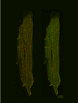
In vivo imaging of brown algal cells in 3D is extremely challenging because of the presence of pigments, such as fucoxanthin and chlorophyll, that diffract light. Moreover, brown algae live in seawater, a high ionic environment that can change the fluorochrome behavior or cause aggregates. Despite the importance of in vivo monitoring the developmental process of brown algal tissues, 4D imaging (x, y, z, t) on a conventional fluorescence microscope is limited. Here, we propose a detailed protocol using a new orange-emitting fluorochrome, styryl benzoindoleninium sulfonate (SBIS), suitable for labeling the plasma membrane of brown algal cells and multicolor in vivo imaging in 3D using confocal and light sheet microscopy. Unlike calcofluor white (CFW), SBIS enables the observation of brown algal cells at thicknesses up to 25 μm and over periods up to 7 days on brown algae such as Ectocarpus sp., Sphacelaria rigidula, and Saccharina latissima. This step-by-step protocol includes labeling of brown algal tissues, mounting for 3D confocal time-lapse microscopy, and mounting for 3D time-lapse light sheet microscopy. The imaging setup and parameters have been optimized for minimizing toxicity for brown algal tissues, improving signal-to-noise ratio, and enabling detailed visualization of cell shape. Therefore, this protocol provides robust and multiplexed imaging with 4D visualization of brown algal cell shape throughout the brown algae growth, offering broad applications to brown algae study at the cellular level.
一种用于在杨树中高效构建大片段缺失的双 gRNA CRISPR/Cas9 系统

A Dual-gRNA CRISPR/Cas9 System for Efficient Generation of Large Fragment Deletions in Poplar

一种用于在杨树中高效构建大片段缺失的双 gRNA CRISPR/Cas9 系统

GY Guoqian Yang
YY Yang Yu
VV Vijaya Kumar Reddy Vulavala
ND Nidhi Dwivedi
CL Chang-Jun Liu
68 Views
Jul 5, 2026
CRISPR/Cas9-based genome editing is a powerful approach for functional genomics and bioenergy research in woody plants. However, conventional single guide RNA (gRNA) strategies predominantly generate small insertions or deletions that may not fully disrupt gene function and often require extensive sequencing for mutation identification. Here, we present an optimized protocol for the efficient generation of large-fragment deletion mutants in Populus tremula × P. alba clone INRA 717-1B4 using a dual-gRNA CRISPR/Cas9 system. Co-expression of two gRNAs flanking the target region induces double-strand breaks at both sites, enabling the deletion of the intervening genomic fragment, typically larger than 50 bp. This protocol describes step-by-step procedures for gRNA design, vector construction, Agrobacterium-mediated transformation, plant regeneration, and molecular validation. Using the PtFBX230 gene as a representative target, large deletions are readily identified by conventional PCR and agarose gel electrophoresis, enabling rapid and cost-effective genotyping. This protocol can be readily adopted to other loci in poplar and related woody species and provides a robust framework for generating null alleles to support functional genomics and bioenergy-related trait engineering in woody plants.
利用酶标仪测定 OsAIM1 的 NADH 依赖性氧化还原酶活性

NADH-Dependent Oxidoreductase Activity Assay of OsAIM1 Using a Microplate Reader

利用酶标仪测定 OsAIM1 的 NADH 依赖性氧化还原酶活性

YH Yuan Hu
SC Song Cui
HL Haishen Li
HH Haigang Hou
ZX Zhuang Xu
BH Benyuan Hao
LC Liang Cai
LZ Liang Zhu
JW Jie Wang
KC Kongyou Chang
WL Wen Li
WS Weihao Shao
SZ Shanshan Zhu
CL Chao Li
ZZ Zhigang Zhao
LJ Ling Jiang
YT Yunlu Tian
XL Xi Liu
SL Shijia Liu
LC Liangming Chen
SZ Shirong Zhou
JW Jianmin Wan
48 Views
Jul 5, 2026
Peroxisomal β-oxidation is a key step in jasmonic acid biosynthesis. Quantitative biochemical characterization of enzymes involved in the β-oxidation pathway is essential for validating their catalytic functions and comparing differences among genetic variants. Existing enzyme activity assays largely rely on chromatographic techniques to quantify substrate consumption or product formation, but these approaches are not well-suited for high-throughput or continuous kinetic measurements. Here, we describe a spectrophotometric assay based on a plate reader determining OsAIM1 enzymatic activity by monitoring the decrease in NADH absorbance at 340 nm. The method employs a 96-well plate reaction system, enabling real-time kinetic measurements and providing a standardized workflow for calculating reaction rates. Reaction components, protein concentration ranges, and data processing parameters were systematically optimized to ensure linearity, reproducibility, and quantitative accuracy. This assay is simple to perform, requires small reaction volumes, and offers relatively high throughput, making it suitable for functional characterization and kinetic analysis of NADH-dependent enzymes.
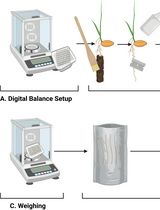
水稻小花开放过程中浆片的分离与形态测量分析

Lodicule Isolation and Morphometric Analysis During Rice Floret Opening

水稻小花开放过程中浆片的分离与形态测量分析

YH Yuan Hu
SC Song Cui
HL Haishen Li
HH Haigang Hou
ZX Zhuang Xu
BH Benyuan Hao
LC Liang Cai
LZ Liang Zhu
JW Jie Wang
KC Kongyou Chang
WL Wen Li
WS Weihao Shao
SZ Shanshan Zhu
CL Chao Li
ZZ Zhigang Zhao
LJ Ling Jiang
YT Yunlu Tian
XL Xi Liu
SL Shijia Liu
LC Liangming Chen
SZ Shirong Zhou
JW Jianmin Wan
35 Views
Jul 5, 2026
Rice lodicules are specialized floral organs located at the base of the ovary that undergo dynamic morphological changes during the flowering period. Water uptake–driven swelling and subsequent dehydration-induced shrinkage of the lodicules trigger floret opening and closure, respectively. Although lodicules play a central role in floret movement, standardized methods for quantitatively monitoring their temporal morphological changes remain limited. Here, we describe a detailed and reproducible workflow for lodicule sampling, dissection, imaging, and quantitative morphometric analysis. Florets are collected at predefined clock time points during the flowering period, and lodicules are carefully isolated under a stereomicroscope. High-resolution imaging is performed under consistent acquisition settings, followed by precise measurement of lodicule length, width, and thickness using image analysis software. This protocol emphasizes positional consistency in sampling, uniform imaging parameters, and standardized data analysis to enhance reproducibility. This method is suitable for evaluating the effects of genetic background or environmental conditions on lodicule morphology. By providing a standardized analytical framework, this protocol enables accurate and quantitative morphometric analysis of rice lodicules during floret opening.

干细胞

将小鼠胚胎干细胞逐步分化为小鼠血管类器官并通过内皮细胞谱系追踪进行质量控制

Stepwise Differentiation of Mouse Embryonic Stem Cells Into Murine Blood Vessel Organoids With Endothelial Lineage Tracing for Quality Control

将小鼠胚胎干细胞逐步分化为小鼠血管类器官并通过内皮细胞谱系追踪进行质量控制

SG Sophie Guelfi
SB Sarah Bopp
GB Gabriele Bergers
138 Views
Jul 5, 2026
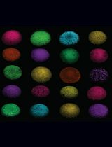
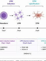
In vitro vascular models are most informative when they recapitulate endothelial assembly within a 3D microenvironment. Blood vessel organoids (BVOs) enable the study of vascular heterogeneity, function, and organ-instructive cues in development, homeostasis, and disease. Here, we present a robust stepwise method to generate murine blood vessel organoids (mBVOs) from feeder-dependent mouse embryonic stem cells (mESCs) of common genetic backgrounds. Embryoid bodies (EBs) are formed using strain-specific seeding densities (day 0–3), followed by mesoderm induction (day 3–6) and vascular induction (day 6–8). Induced EBs are embedded in collagen I with Geltrex to drive sprouting and network formation (day 8–13). Vascular networks are microdissected and grown in suspension to yield mature mBVOs (day 21–30). The inclusion of a Cre-inducible VE-cadherin-GFP reporter line enables a quantitative quality control, reducing variability by excluding poorly differentiated organoids. The protocol reliably produces ~100 mBVOs per differentiation and is compatible with engineered mouse strains for gain- and loss-of-function studies, functional assays of vascular plasticity, and syngeneic grafting to assess perfusion. Thus, mBVOs provide a scalable and traceable 3D platform that bridges endothelial assays, mouse models, and human organoid systems.