

现期刊物2026
卷册: 16, 期号: 14
生物化学
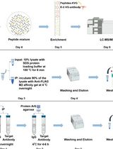
A Practical Experimental Protocol for Identification and Validation of UFMylation Substrate in Human Cells
人细胞中UFM1修饰底物的鉴定与验证实验方案
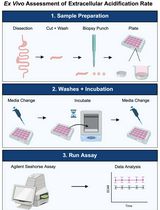
Ex Vivo Assessment of Extracellular Acidification Rate in Murine Intestinal Tissue
小鼠肠道组织细胞外酸化率的离体测定方法
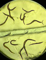
Iodine Staining of Glycogen Storage in Caenorhabditis elegans
秀丽隐杆线虫糖原储存的碘染色检测方法
生物信息学与计算生物学
Actin Quantification Using the Filamentous Actin Segmentation Tool (FAST)
利用丝状肌动蛋白分割工具(FAST)定量分析肌动蛋白
生物工程
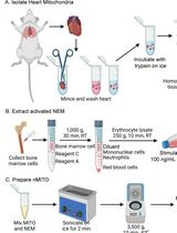
Preparation and Characterization of Neutrophil Membrane-Fused Mitochondria (nMITO)
中性粒细胞膜融合线粒体(nMITO)的制备与表征
癌症生物学
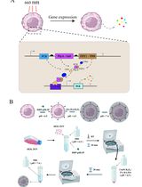
Light-Regulated Cancer Immunotherapy Using Individually Encapsulated Synthetic Circuit–Engineered Cells
利用单细胞封装的合成回路工程细胞实现光控癌症免疫治疗
细胞生物学
A Universal Resazurin-Based Viability Assay for Prokaryotic and Eukaryotic Cells in 2D and 3D Cultures
适用于二维和三维培养原核与真核细胞的通用刃天青活力检测方法


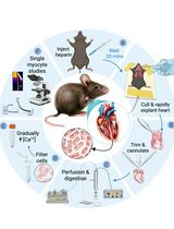
A Simplified Langendorff-Based Method for Mouse Cardiac Myocyte Isolation
基于Langendorff灌流的小鼠心肌细胞简化分离方法
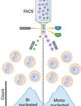
Isolation of Mononucleated and Binucleated Hepatocytes by Flow Cytometry
利用流式细胞术分离单核与双核肝细胞
发育生物学
In Vivo Light-Sheet Imaging of Senescence Reporter Activity in a Transparent Killifish
利用光片显微镜活体观察透明非洲青鳉中的衰老报告基因活性
免疫学
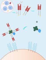
A Streamlined and Time-Saving Approach to Generate HLA-DR15 MHC Class II Tetramers via In Vivo Biotinylation
通过体内生物素化简便快速制备HLA-DR15 MHC II类四聚体
微生物学
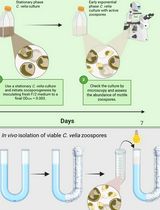
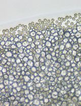
Separating Chromera velia Zoospores From Culture and Estimating Their Average Motility Speed and Lifespan
从培养物中分离Chromera velia游动孢子并测定其平均运动速度和寿命


Optimized Field Collection and Gut Dissection Workflows for Microbiome Studies of the Citrus Root Weevil, Diaprepes abbreviatus
Diaprepes abbreviatus 微生物组研究的野外采集与肠道解剖流程优化
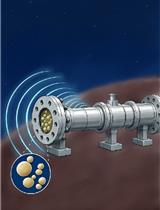
Assessment of Saccharomyces cerevisiae Survival Upon Exposure to Transient High Pressure and Temperature in a High-Intensity Shock Tube for Astrobiology (HISTA)
利用天体生物学高强度激波管(HISTA)评估酿酒酵母在瞬时高压高温条件下的存活情况
分子生物学
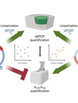
An Accurate and Precise ddPCR-Based Method for Determining the Concentration of Plasmid DNA
基于ddPCR准确测定质粒DNA浓度的高精度方法
神经科学
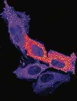
Measuring PINK1 Activity in Single Cells Using a PINK1 Kinase Activity Reporter
利用PINK1激酶活性报告器检测单细胞PINK1活性
植物科学
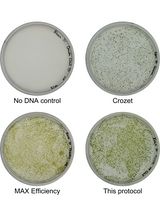
Simple Electroporation of Chlamydomonas reinhardtii Strains With an Intact Cell Wall
完整细胞壁莱茵衣藻藻株的简易电转化方法

Gene Editing in Chlamydomonas Using the SCREAM Technique
利用SCREAM技术开展衣藻基因编辑
干细胞
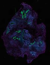
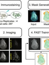
Histological Processing of Organoids for Immunostaining
类器官免疫染色的组织学处理方法
Satellite Cell Isolation, Culture, and Infection After Retroviral Preparation
逆转录病毒制备后卫星细胞的分离培养与感染
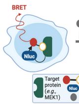
Protocol for Measuring Drug–Target Engagement in Mouse Colorectal Cancer Organoids Using NanoBRET Assay
利用NanoBRET检测小鼠结直肠癌类器官中药物与靶标结合的实验方法