往期刊物2025

卷册: 15, 期号: 8

left

Oct

20

Oct

5

Sep

20

Sep

5

Aug

20

Aug

5

Jul

20

Jul

5

Jun

20

Jun

5

May

20

May

5

Apr

20

Apr

5

Mar

20

Mar

5

Feb

20

Feb

5

Jan

20

Jan

5

right

生物化学

一种使用 SpyDock 修饰树脂的稳定且简便的蛋白质纯化方法

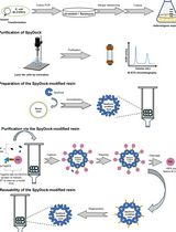
A Robust and Easy Protein Purification Method Using SpyDock-Modified Resin

一种使用 SpyDock 修饰树脂的稳定且简便的蛋白质纯化方法

XY Xiaofeng Yang
ZL Zhanglin Lin
YX Ya Xiang
BC Binrui Chen
ZL Zisha Lao
1511 Views
Apr 20, 2025
Protein purification is a critical step in both life sciences and biomanufacturing. Traditional affinity chromatography (AC) methods, including His-tag-based purification, provide high-purity proteins but are limited by the high cost of resins and the need for additional tag-removal steps. In this protocol, we present a reusable SpyDock-modified epoxy resin coupled with a pH-inducible self-cleaving intein for direct purification of proteins with authentic N-termini. This method enables efficient protein purification from cell lysates, achieving high purity (>90%) and yields comparable to the His-tag approach, without requiring tag removal. The SpyDock-modified resin protocol is robust, easy to implement, and cost-effective, making it suitable for both research and large-scale industrial applications.
使用 Spy Chemistry 进行抗体纯化

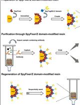
Antibody Purification Using Spy Chemistry

使用 Spy Chemistry 进行抗体纯化

XY Xiaofeng Yang
ZL Zhanglin Lin
YX Ya Xiang
ZL Zisha Lao
1255 Views
Apr 20, 2025
Antibody purification is a fundamental technology for therapeutic and diagnostic applications. While traditional methods like ammonium sulfate precipitation and polyethylene glycol precipitation are cost-effective, they often result in low purity and require multiple purification steps. Protein A–based affinity chromatography, the gold standard for antibody purification, provides high specificity but can be further improved to increase loading capacity and reduce costs. In this protocol, we introduce a novel approach for purifying high-quality, high-purity antibodies from complex samples using SpyFixer/Z domain–modified resin. This method utilizes Spy chemistry for site-specific immobilization of the Z domain of Protein A, significantly enhancing antibody loading capacity up to 200 mg/mL resin and ensuring stable, durable immobilization. Using this protocol, we achieved >90% purity of human immunoglobulin G (hIgG) from diverse sources, including E. coli cell lysates, human serum, and industrial fermentation broth. The SpyFixer/Z domain–modified resin protocol is simple, cost-effective, and offers a robust, scalable solution for efficient antibody purification in bioengineering applications. This immobilization scheme based on Spy chemistry can also be extended to other immunoglobulin-binding proteins, such as Protein G and Protein L, to develop high-efficiency affinity resins.
基于Azo-Xylan及其变体的Endo-1,4-β-D-木聚糖酶活性检测方法

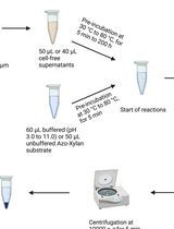
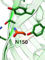
Endo-1,4-β-D-xylanase Assay Using Azo-Xylan and Variants Thereof

基于Azo-Xylan及其变体的Endo-1,4-β-D-木聚糖酶活性检测方法

LB Luca Bombardi
AC Annalaura Coltro
SF Salvatore Fusco
1225 Views
Apr 20, 2025
Xylan is the main component of hemicellulose and consists of a complex heteropolysaccharide with a heterogeneous structure. This framework, in addition to the crystalline structure of cellulosic fibers and the rigidity of lignin, makes lignocellulosic biomass (LCB) highly recalcitrant to degradation. Xylanases are glycoside hydrolases that cleave the β-1,4-glycoside linkages in the xylan backbone and have attracted increasing attention due to their potential uses in various industrial sectors such as pulp and paper, baking, pharmaceuticals, and lignocellulosic biorefining. For decades, the measurement of xylanase activity was based on reducing sugar quantification methods like DNS or Nelson/Somogyi assays, with numerous limitations in terms of specificity and interference from other enzymatic activities. A better alternative is the colorimetric Azo-Xylan assay, which specifically measures the endo-1,4-β-D-xylanase activity. In this study, the Azo-Xylan protocol was adapted from the company Megazyme to determine the enzymatic activity of thermostable xylanases produced by microbial consortia (i.e., microbiomes), aiming to determine biochemical features such as temperature and pH optima, thermostability, and shelf life. This modified approach offers a rapid, cost-effective, and highly specific method for the determination of xylanase activity in complex mixtures, helping the development of a xylanase-based method for the hydrolysis of hard-degrading substrates in bio-based industries.

生物信息学与计算生物学

适用于复杂群体结构的多样性群体基因定位的GWAS分析流程

GWAS Procedures for Gene Mapping in Diverse Populations With Complex Structures

适用于复杂群体结构的多样性群体基因定位的GWAS分析流程

ZZ Zhen Zuo
ML Mingliang Li
DL Defu Liu
QL Qi Li
BH Bin Huang
GY Guanshi Ye
JW Jiabo Wang
YT You Tang
Zhiwu Zhang Zhiwu Zhang
2256 Views
Apr 20, 2025
With reduced genotyping costs, genome-wide association studies (GWAS) face more challenges in diverse populations with complex structures to map genes of interest. The complex structure demands sophisticated statistical models, and increased marker density and population size require efficient computing tools. Many statistical models and computing tools have been developed with varied properties in statistical power, computing efficiency, and user-friendly accessibility. Some statistical models were developed with dedicated computing tools, such as efficient mixed model analysis (EMMA), multiple loci mixed model (MLMM), fixed and random model circulating probability unification (FarmCPU), and Bayesian-information and linkage-disequilibrium iteratively nested keyway (BLINK). However, there are computing tools (e.g., GAPIT) that implement multiple statistical models, retain a constant user interface, and maintain enhancement on input data and result interpretation. In this study, we developed a protocol utilizing a minimal set of software tools (BEAGLE, BLINK, and GAPIT) to perform a variety of analyses including file format conversion, missing genotype imputation, GWAS, and interpretation of input data and outcome results. We demonstrated the protocol by reanalyzing data from the Rice 3000 Genomes Project and highlighting advancements in GWAS model development.
基于MrBayes的贝叶斯系统发育分析全流程方案:从序列比对到模型选择与系统发育推断

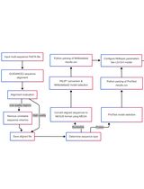
A Comprehensive Protocol for Bayesian Phylogenetic Analysis Using MrBayes: From Sequence Alignment to Model Selection and Phylogenetic Inference

基于MrBayes的贝叶斯系统发育分析全流程方案:从序列比对到模型选择与系统发育推断

JW Jinxing Wang
FC Fangmin Chen
XX Xu Xiao
XY Xinyao Yang
WX Wanting Xia
1223 Views
Apr 20, 2025
Bayesian phylogenetic analysis is essential for elucidating evolutionary relationships among organisms. Traditional methods often rely on fixed models and manual parameter settings, which can limit accuracy and efficiency. This protocol presents an integrated workflow that leverages GUIDANCE2 for rigorous sequence alignment, ProtTest and MrModeltest for robust model selection, and MrBayes for phylogenetic tree estimation through Bayesian inference. By automating key steps and providing detailed command-line instructions, this protocol enhances the reliability and reproducibility of phylogenetic studies.

生物物理学

结合X射线光子相关光谱、显微成像与荧光漂白恢复技术研究朊蛋白凝聚体的相分离及液-固转变过程

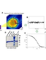
X-Ray Photon Correlation Spectroscopy, Microscopy, and Fluorescence Recovery After Photobleaching to Study Phase Separation and Liquid-to-Solid Transition of Prion Protein Condensates

结合X射线光子相关光谱、显微成像与荧光漂白恢复技术研究朊蛋白凝聚体的相分离及液-固转变过程

Mariana J. do Amaral Mariana J. do Amaral
AP Aline R. Passos
SM Satabdee Mohapatra
Maria Heloisa Freire Maria Heloisa Freire
SW Susanne Wegmann
YC Yraima Cordeiro
2177 Views
Apr 20, 2025
Biomolecular condensates are macromolecular assemblies constituted of proteins that possess intrinsically disordered regions and RNA-binding ability together with nucleic acids. These compartments formed via liquid-liquid phase separation (LLPS) provide spatiotemporal control of crucial cellular processes such as RNA metabolism. The liquid-like state is dynamic and reversible, containing highly diffusible molecules, whereas gel, glass, and solid phases might not be reversible due to the strong intermolecular crosslinks. Neurodegeneration-associated proteins such as the prion protein (PrP) and Tau form liquid-like condensates that transition to gel- or solid-like structures upon genetic mutations and/or persistent cellular stress. Mounting evidence suggests that progression to a less dynamic state underlies the formation of neurotoxic aggregates. Understanding the dynamics of proteins and biomolecules in condensates by measuring their movement in different timescales is indispensable to characterize their material state and assess the kinetics of LLPS. Herein, we describe protein expression in E. coli and purification of full-length mouse recombinant PrP, our in vitro experimental system. Then, we describe a systematic method to analyze the dynamics of protein condensates by X-ray photon correlation spectroscopy (XPCS). We also present fluorescence recovery after photobleaching (FRAP)-optimized protocols to characterize condensates, including in cells. Next, we detail strategies for using fluorescence microscopy to give insights into the folding state of proteins in condensates. Phase-separated systems display non-equilibrium behavior with length scales ranging from nanometers to microns and timescales from microseconds to minutes. XPCS experiments provide unique insights into biomolecular dynamics and condensate fluidity. Using the combination of the three strategies detailed herein enables robust characterization of the biophysical properties and the nature of protein phase-separated states.

癌症生物学

小鼠原代周细胞的分离和培养

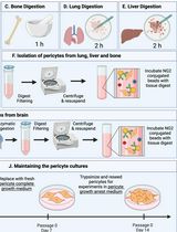
Isolation and Culture of Primary Pericytes from Mouse

小鼠原代周细胞的分离和培养

TM Tamara McErlain
CB Cristina M. Branco
MM Meera Murgai
2115 Views
Apr 20, 2025
Pericytes are essential for tissue homeostasis, functioning to regulate capillary blood flow. Dysfunctional pericytes are implicated in various pathologies, including cancer progression. Despite their important function in both health and disease, pericytes remain understudied due to a lack of robust model systems that accurately reflect their in vivo biology. Here, we present a comprehensive protocol for isolating and culturing primary pericytes from murine lung, brain, bone, and liver tissues, based on NG2 expression using an antibody-conjugated magnetic bead approach. Our protocol emphasizes the importance of physiological oxygen tension during ex vivo culture (10% O2 for lung pericytes and 5% O2 for brain, bone, and liver pericytes). These conditions stabilize the expression of characteristic pericyte markers at both the transcriptional and protein levels. Importantly, we optimized growth conditions to limit the expression of the plasticity factor Klf4 in order to prevent spontaneous phenotypic switching in vitro. This protocol provides a reliable and reproducible method for obtaining pericytes suitable for high-throughput analyses in order to explore pericyte biology in both physiological and pathological contexts.

细胞生物学

利用三功能连接子标记的鬼笔环肽结合扩增显微术可视化F-Actin

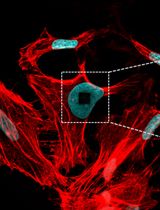
Visualization of F-Actin Through Expansion Microscopy (ExM) with Trifunctional Linker-Conjugated Phalloidin

利用三功能连接子标记的鬼笔环肽结合扩增显微术可视化F-Actin

JH Jianjun Huang
GW Gang Wen
TI Thibo Iven
DL Débora Linhares
LK Leewon Koo
MS Markus Sauer
WD Wim Dehaen
VL Volker Leen
JH Johan Hofkens
1451 Views
Apr 20, 2025
Expansion microscopy (ExM) is an imaging technique that enables super-resolution imaging of biological specimens using conventional confocal microscopy. This process entails the isotropic physical expansion of a (biomolecular) sample that has been cross-linked to a swellable polymer. The grafting of biomolecules (and the subsequent fluorescent readout) is accomplished by introducing an acryloyl group to the amine groups of lysine residues within the proteins, enabling subsequent imaging. However, visualizing actin filaments with high spatial resolution using ExM remains challenging. Herein, we report the construction of a phalloidin conjugate containing actin stains and their application in ExM. This protocol highlights the efficacy of trifunctional linker (TRITON/Actin-ExM) for F-actin imaging, demonstrating that TRITON-labeled actin allows for efficient anchoring and signal retention, enabling robust visualization of actin filaments in expansion microscopy.
用于可视化与定量骨细胞腔道系统(LCS)的新型优化硝酸银染色方法

A Novel Optimized Silver Nitrate Staining Method for Visualizing and Quantifying the Osteocyte Lacuno-Canalicular System (LCS)

用于可视化与定量骨细胞腔道系统(LCS)的新型优化硝酸银染色方法

JW Jinlian Wu
CX Chunchun Xue
QL Qiang Li
HW Hongjin Wu
JZ Jie Zhang
CW Chenglong Wang
WD Weiwei Dai
LW Libo Wang
850 Views
Apr 20, 2025
The osteocyte lacuno-canalicular system (LCS) plays a crucial role in maintaining bone homeostasis and mediating cellular mechanotransduction. Current histological techniques, particularly the Ploton silver nitrate staining method, face challenges such as variations in solution concentrations and types as well as a lack of standardization, which limits their broader application in osteocyte research. In this study, we present a simplified and more effective silver nitrate staining protocol designed to address these issues. Our method utilizes a 1 mol/L silver nitrate solution combined with optimized gelatin-formic acid solutions at varying concentrations (0.05%–0.5% type-B gelatin and 0.05%–5% formic acid, or 1%–2% type-B gelatin and 0.1%–2% formic acid). Staining is performed for 1 h under 254 nm ultraviolet light or 90 min under room light, followed by washing with Milli-Q water to terminate staining. This novel optimized method yields consistent and distinct staining of the osteocyte LCS across multiple species, demonstrating superior efficiency and reliability compared to the Ploton method. It will significantly advance research in osteocyte biology and provide a valuable tool for exploring the adaptive evolution of osteocyte LCS morphology and function across various taxa.

免疫学

体外诱导骨髓来源树突状细胞(BMDC)用于抗原呈递实验

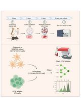
In Vitro Bone Marrow–Derived Dendritic Cells (BMDC) Generation for Antigen Presentation Assay

体外诱导骨髓来源树突状细胞(BMDC)用于抗原呈递实验

SS Sudhakar Singh
Azeez Tehseen Azeez Tehseen
MI Mohammed Shaaz Iqbal
SS Sharvan Sehrawat
2663 Views
Apr 20, 2025
Dendritic cells (DC) are sentinel cells of the immune system that process and present antigens to activate T cells, thus serving to bridge the innate and adaptive immune systems. DCs are particularly efficient at cross-presentation whereby exogenously acquired antigens are processed and presented in context with MHCI molecules to activate CD8+ T cells. Assaying antigen presentation by DCs is a critical parameter in assessing immune functionality. However, the low abundance of bona fide DCs within the lymphoid compartments limits the utility of such assays. An alternative approach employing the culturing of bone marrow cells in the presence of factors needed for DC lineage commitment can result in the differentiation of bone marrow dendritic cells (BMDCs). This protocol details the process of in vitro generation of BMDCs and demonstrates their subsequent utility in antigen presentation assays. The protocol described can be adapted to various conditions and antigens.

微生物学

基于扩展型CPER的快速无质粒重组正链RNA病毒构建方法:适用于IRES介导翻译系统

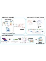
Rapid Plasmid-Free Generation of Recombinant Positive-Strand RNA Viruses That Use IRES-Mediated Translation Using an Expansion of the Circular Polymerase Extension Reaction (CPER)

基于扩展型CPER的快速无质粒重组正链RNA病毒构建方法:适用于IRES介导翻译系统

HY Hirotaka Yamamoto
TT Tomokazu Tamura
TF Takausuke Fukuhara
1173 Views
Apr 20, 2025
Reverse genetics systems in virology are technologies used to generate recombinant viruses, enabling the manipulation of viral genes. Recombinant viruses facilitate the investigation of pathogenesis and the development of antivirals. In studies of positive-sense single-stranded RNA (ssRNA) viruses, a reverse genetics approach typically uses infectious viral cDNA clones derived from bacterial artificial chromosomes and plasmids or from the in vitro ligation of viral cDNA fragments. However, these methods are time-consuming, involve complex procedures, and do not always successfully generate recombinant viruses. Possible reasons for unsuccessful outcomes include i) viral sequences exhibiting toxicity in bacterial systems, ii) the duplication of viral genes observed in some strains, complicating the acquisition of correct cDNA clones, and iii) certain cell lines being highly susceptible to infection but difficult to transfect with nucleotides. For these reasons, a simple and rapid reverse genetics system is needed to accelerate research on ssRNA viruses. The circular polymerase extension reaction (CPER) method offers a solution by eliminating the need for molecular cloning in bacteria, enabling the generation of recombinant viruses over a shorter timeframe. This method has been widely adopted for the study of ssRNA viruses, including SARS-CoV-2 and flaviviruses. Recently, we expanded the CPER method for ssRNA viruses using internal ribosome entry site (IRES)-mediated translation. This protocol details the experimental procedures, using bovine viral diarrhea virus as an example—one of the most challenging viruses for generating viral cDNA clones because of the factors listed above.
基于Intein的生物传感器用于体内蛋白稳定性监测

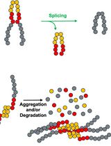
Monitoring Protein Stability In Vivo Using an Intein-Based Biosensor

基于Intein的生物传感器用于体内蛋白稳定性监测

JS John S. Smetana
TA Tia M. Ariagno
AS Ahyun Son
SH Scott Horowitz
CL Christopher W. Lennon
1046 Views
Apr 20, 2025
Inteins are elements translated within host proteins and removed via a unique protein splicing reaction. In this process, the two peptide bonds flanking the intein are rearranged, releasing the intein and leaving a standard peptide bond in its place. Due to their ability to shuffle peptide bonds in a specific and controlled manner, inteins have proven valuable in protein engineering, leading to the development of numerous impactful technologies. In one application, intein-based biosensors link the activity of a host protein to intein excision. Recently, we developed a biosensor to measure protein stability in vivo, in which the removal of an intein-protein fusion is required for antibiotic resistance. In our protocol, cells expressing our biosensor are logarithmically diluted and spotted on agar plates containing increasing levels of antibiotics. Following incubation, quantitative survival curves can be generated. We also developed a dual protein stability sensor where both antibiotic resistance and fluorescence can be used as readouts and demonstrated that co-expression of the chaperonin GroEL can promote survival and fluorescence. Taken together, our novel intein-based biosensor adds to the available tools to measure protein stability within the cellular environment.
适用于PCR微生物DNA筛查的可扩展碱提取方法

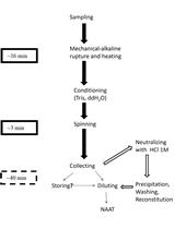
Scalable Alkaline Extraction Protocol for Microbial DNA Screening by PCR

适用于PCR微生物DNA筛查的可扩展碱提取方法

SG Stavroula Goudoudaki
MK Manousos E. Kambouris
SK Stavroula Kritikou
AM Afroditi Milioni
AV Aristea Velegraki
YM Yiannis Manoussopoulos
GP George P. Patrinos
1010 Views
Apr 20, 2025
In molecular diagnosis, DNA extraction kits are sample-specific and proprietary, preventing lateral distribution among similar facilities from different sectors to alleviate supply shortages during a crisis. Previous fast extraction protocols such as detergent-based ones allow fast DNA extraction for nucleic acid amplification tests (NAAT), mainly polymerase chain reaction (PCR). The use of NaOH (dense alkali) to rupture cells and nuclei and destabilize the conformation of DNases might alleviate shortages and costs while retaining enough robustness to treat complicated samples with minimal environmental and logistical footprint. Biological samples are hand-crushed using a pestle in 1.5 mL tubes with 360 μL of 0.2 M NaOH for 3–5 min and incubated at 75 °C for 10 min. For immediate use, 115.2 μL of 1 M Tris (pH 8) and 364.8 μL nuclease-free water are added, and the sample is vortexed for 10 s and spun at 10,000× g for 3 min; then, 700 μL is transferred to a clean microtube. Two serial dilutions follow, and all concentrations are used as templates for PCR. A refined, storable extract can be produced by adding 70 μL of HCl 1 M (instead of Tris-HCl) and one volume of cold isopropanol to the extract for standard precipitation. This method can increase throughput in emergencies by field deployment in resource-limited settings (RLS) or allow benchtop backup in cases of acquisition disruption or sample surge in established facilities. The crude extract can be used for immediate PCR in both benchtop and portable thermocyclers, thus allowing NAAT in resource-limited settings with low costs and waste footprint or during prolonged crises, where supply chain failures may occur. The refined version produces alcohol-precipitated nucleic acids, suitable for both immediate use and for storage or dispatch for spatiotemporally separate analysis while offering much better amplification quality with a small increase in time and minimal increase in expendables/chemicals needed.
基于微型Percoll密度梯度的酵母静息细胞分离方法

A Miniaturized Percoll Gradient Method for Isolation of Quiescent Cells of Yeast

基于微型Percoll密度梯度的酵母静息细胞分离方法

CZ Cenxuan Zu
JD Jia Dong
YZ Yue Zhang
YZ Yao Zhang
ZH Zongzheng Huang
Shaolan Zou Shaolan Zou
878 Views
Apr 20, 2025
Quiescence, the temporary and reversible exit from proliferative growth, is a fundamental biological process. Budding yeast is a preeminent model for studying cellular quiescence owing to its rich experimental toolboxes and evolutionary conservation across eukaryotic pathways and processes that control quiescence. Yeast quiescent cells are reported to be isolated by the continuous linear Percoll gradient method and identified by combining different features such as cell cycle, heat resistance, and cell morphology (single cell). Generally, 10–25 mL of Percoll isotonic solution is first obtained by mixing Percoll with NaCl in 12.5–30 mL centrifugal tubes. Then, the gradient is prepared at high speed for 15–60 min. Finally, approximately 2 × 10⁹ cells are collected, overlaid onto the preformed gradient, and centrifuged to obtain distinct cell fractions. This method requires more reagents and samples and special centrifuges and centrifuge tubes. Besides the cost, it is less favorable for experiments that require high-throughput analyses with a small volume of sample each time. The protocol described here aims to solve those problems by combining the use of 2 mL centrifugal tubes with density marker beads. The protocol also focuses on how to optimize the buoyant density distribution of the density gradient solution such that the density bands better match those of different fraction cells. This will help fully separate quiescent and non-quiescent cells. The protocol can be easily adapted to a wide variety of unicellular microbes with different buoyancy density differentiation during cultivation, such as yeast and bacteria.

神经科学

线虫PVD神经元激光损伤后树突与轴突功能性再生的分析与定量

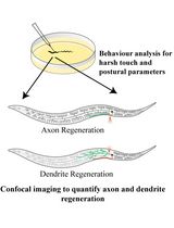
Analysis and Quantification of Functional Regeneration of Dendrite and Axon of PVD Neuron After Laser Injury in Caenorhabditis elegans

线虫PVD神经元激光损伤后树突与轴突功能性再生的分析与定量

HB Harjot Kaur Brar
DB Dikshalee Bassi
AG Anindya Ghosh-Roy
1633 Views
Apr 20, 2025
Research into nervous system injuries and regeneration has emerged as a crucial field of study. In many cases such as trauma or stroke, both axons and dendrites are equally damaged; however, studying injury and repair mechanisms in both neurite processes (axons and dendrites) of the same neuron has been challenging. Additionally, correlating the behavioral aspects of neuronal injury with anatomical regeneration is important for a better understanding of the functional rewiring process. Here, we describe protocols for injuring the dendrites and the axon of the PVD neuron of C. elegans using a two-photon infrared (IR) femtosecond laser system, and subsequent imaging of injured neurites during the course of regeneration. Additionally, we describe the protocols for the behavioral study concerning the PVD neuron and their analysis, which can offer valuable insights. These assays can be implemented to assess the function of the pathways that play specific roles in dendrite vs. axon regeneration.
小鼠社会虚弱指数(mSFI):标准化实验方案

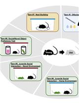
The Mouse Social Frailty Index (mSFI): A Standardized Protocol

小鼠社会虚弱指数(mSFI):标准化实验方案

CC Charles W. Collinge
MR Maria Razzoli
AB Alessandro Bartolomucci
1121 Views
Apr 20, 2025
The advent of geroscience engendered the development of approaches to quantify the aging process and estimate biological age on an individual level. Recognizing that declines observed in aging are not only physical but also social led us to develop a mouse Social Frailty Index (mSFI) designed to quantify age-related impairments of social functioning in mice. The mSFI consists of seven behavioral assays that measure essential facets of social behavioral functioning in mice: social communication, social interaction, and social functional ability. The assays that comprise the mSFI are all minimally disruptive, relatively simple to execute, and optimized for compatibility with longitudinal studies utilizing experimental interventions relevant to geroscience. The mSFI is conducted over AM and PM sessions spanning a maximum of 3.5 days, using materials common to most animal facilities. The data for all assays is obtained observationally, manually recorded, and entered into predefined template sheets that automate the computation of the mSFI. We have demonstrated the validity and applicability of the mSFI across multiple laboratory sites and experiments. This index has proven to discriminate between differential trajectories of biological aging driven by sex, progeria, or social stress-relevant contexts. The mSFI represents a novel index to quantify trajectories of biological aging in mice, and its application may help elucidate the social dimensions of the aging process.

植物科学

活体植物根部细胞核的近红外自发荧光成像

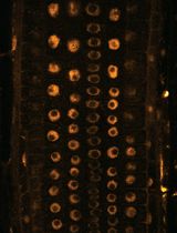
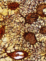
Near-Infrared Autofluorescence Imaging of Nuclei in Living Plant Roots

活体植物根部细胞核的近红外自发荧光成像

AY Akira Yoshinari
MN Masayoshi Nakamura
1286 Views
Apr 20, 2025
In live-cell imaging, autofluorescence is often regarded as a negative factor that interferes with the accurate visualization of target fluorescence due to a phenomenon known as crosstalk. However, autofluorescence has also been effectively utilized as an organellar marker. For instance, the intense autofluorescence of chlorophyll in the red wavelength is widely used to visualize chloroplasts, the photosynthetic organelle in plants. Recently, we demonstrated that nuclei in plant cells emit phytochrome-derived autofluorescence in the red to infrared wavelength range, which can be visualized by a conventional confocal microscope equipped with a 640 nm laser. Here, we present protocols for growing plants and conducting confocal imaging of the near-infrared autofluorescence of nuclei in Arabidopsis thaliana.
植物样本中阴离子磷脂所有分子种类与类别的同步检测及半定量新方法

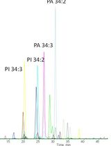
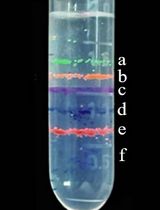
A New Approach to Detect and Semi-quantify All Molecular Species and Classes of Anionic Phospholipids Simultaneously in Plant Samples

植物样本中阴离子磷脂所有分子种类与类别的同步检测及半定量新方法

MG Manon Genva
CM Cécile Mirande-Bret
LF Laetitia Fouillen
1200 Views
Apr 20, 2025
Membranes are very complex and dynamic structures that are essential for plant cellular functions and whose lipidic composition can be influenced by numerous factors. Anionic phospholipids, which include phosphatidylserine, phosphatidic acid, phosphatidylinositol, and phosphoinositides are key components of these membranes as they are involved in plant cell signaling and as even slight modifications in their quantities may largely impact the cell metabolism. However, the presence of these compounds in low amounts, as well as their poor stability during analysis by mass spectrometry, make their study very complicated. In addition, the precise quantification of all anionic phospholipid species is not possible by lipid separation using thin-layer chromatography followed by the analysis of their fatty acyl chains by gas chromatography. Here, we describe a straightforward strategy for the extraction and semi-quantification of all anionic phospholipid species from plant samples. Our method is based on the derivatization of the anionic phospholipids, and more especially on their methylation using trimethylsilyldiazomethane, followed by analysis by high-performance liquid chromatography coupled with a triple quadrupole mass spectrometer. This approach allows largely improving the sensitivity of the analysis of anionic phospholipids from plant samples, which will help to gain deeper insights into the functions and dynamics of these key parts of plant cellular signaling.
基于TMV-GFP系统的植物病原菌候选效应子功能筛选流程

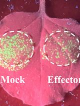
Workflow for a Functional Assay of Candidate Effectors From Phytopathogens Using a TMV-GFP-based System

基于TMV-GFP系统的植物病原菌候选效应子功能筛选流程

PC Peng Cao
HS Haotian Shi
JC Jialan Chen
LC Langjun Cui
MZ Meixiang Zhang
YA Yuyan An
1052 Views
Apr 20, 2025
The ability to efficiently screen plant pathogen effectors is crucial for understanding plant–pathogen interactions and developing disease-resistant crops. Traditional methods are often labor-intensive and time-consuming. Here, we present a robust, high-throughput screening assay using the tobacco mosaic virus–green fluorescent protein (TMV-GFP) vector system. The screening system combines the TMV-GFP vector and Agrobacterium-mediated transient expression in the model plant Nicotiana benthamiana. This system enables the rapid identification of effectors that interfere with plant immunity (both activation and suppression). The biological function of these effectors can be easily evaluated within six days by observing the GFP fluorescence signal using a UV lamp. This protocol significantly reduces the time required for screening and increases the throughput, making it suitable for large-scale studies. The method is versatile, cost-effective, and can be adapted to effectors with immune interference activity from various pathogens.

干细胞

人骨骼肌干细胞细胞核的三维可视化集成流程

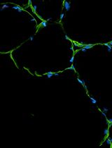
An Integrated Workflow for Three-Dimensional Visualization of Human Skeletal Muscle Stem Cell Nuclei

人骨骼肌干细胞细胞核的三维可视化集成流程

JP Jeremy R. Pearson
NM Noraida Martinez-Rivera
IT Irma Torres-Vasquez
PG Philip M. Gallagher
ER Eduardo Rosa-Molinar
1616 Views
Apr 20, 2025
Skeletal muscle–specific stem cells are responsible for regenerating damaged muscle tissue following strenuous physical activity. These muscle stem cells, also known as satellite cells (SCs), can activate, proliferate, and differentiate to form new skeletal muscle cells. SCs can be identified and visualized utilizing optical and electron microscopy techniques. However, studies identifying SCs using fluorescent imaging techniques vary significantly within their methodology and lack fundamental aspects of the guidelines for rigor and reproducibility that must be included within immunohistochemical studies. Therefore, a standardized method for identifying human skeletal muscle stem cells is warranted, which will improve the reproducibility of future studies investigating satellite activity. Additionally, although it has been suggested that SC shape can change after exercise, there are currently no methods for examining SC morphology. Thus, we present an integrated workflow for three-dimensional visualization of satellite cell nuclei, validated by the spatial context of the fluorescent labeling and multichannel signal overlap. Our protocol includes, from start to finish, post-biopsy extraction and embedding, tissue sectioning, immunofluorescence, imaging steps and acquisition, and three-dimensional data post-processing. Because of the depth volume generated from the confocal microscope z-stacks, this will allow future studies to investigate the morphology of SC nuclei and their activity, instead of traditionally observing them in two-dimensional space (x, y).

更正

校正:快速且完全的蛋白质生物共轭的四嗪氨基酸编码技术

Correction Notice: Tetrazine Amino Acid Encoding for Rapid and Complete Protein Bioconjugation

校正:快速且完全的蛋白质生物共轭的四嗪氨基酸编码技术

AE Alex J. Eddins
AP Abigail H. Pung
RC Richard B. Cooley
RM Ryan A. Mehl
324 Views
Apr 20, 2025