

杂志社论


综述
RNA Detection Technologies: A Method‑Centric Guide to Principles and Reproducibility
RNA检测技术:原理解析与可重复性实践
生物化学
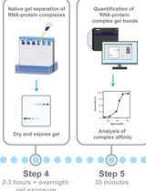
Electrophoretic Mobility Shift Assay (EMSA) for Assessing RNA–Protein Binding and Complex Formation Using Recombinant RNA-Binding Proteins and In Vitro–Transcribed RNA
利用重组 RNA 结合蛋白和体外转录 RNA,通过 EMSA 评估 RNA–蛋白质结合及复合物形成

Simultaneous Immunofluorescence-Based In Situ mRNA Expression and Protein Detection in Bone Marrow Biopsy Samples
基于免疫荧光的骨髓活检样本原位 mRNA 表达与蛋白同步检测
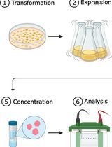
One-Step Affinity Purification of MarathonRT Reverse Transcriptase for RNA Sequencing Applications
用于 RNA 测序的 MarathonRT 逆转录酶一步亲和纯化方法
生物信息学与计算生物学
Enhanced RNA-Seq Expression Profiling and Functional Enrichment in Non-model Organisms Using Custom Annotations
基于自定义注释的非模式生物 RNA-seq 表达谱与功能富集分析优化
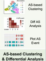
Stepwise Protocol for Alternative Splicing Analysis in Single-Cell SMART-Seq2 RNA-Seq Data
单细胞 SMART-Seq2 RNA-seq 数据中可变剪接分析的分步流程
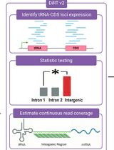
DiRT v2.0: An Optimized Pipeline for Detecting Dicistronic tRNA-mRNA Transcripts in Plants
DiRT v2.0:用于检测植物双顺反子 tRNA-mRNA 转录本的优化流程
免疫学
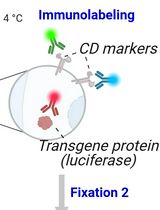
PrimeFlowTM Assay for Cell Type–Specific Co-detection of Transgene RNA and Protein in Mouse Spleens From Preclinical Studies
基于 PrimeFlowTM 的临床前小鼠脾脏样本转基因 RNA 与蛋白细胞类型特异性同步检测
微生物学
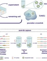
High-Resolution Mapping of RNA–RNA Interactions Across the HIV-1 Genome With HicapR
基于 HiCapR 的 HIV-1 全基因组 RNA–RNA 相互作用高分辨率图谱构建
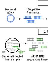
Enriching Bacteria-Specific RNA From Host Samples Before NGS With Transcript-Capture
基于转录本捕获的宿主样本细菌特异性 RNA 富集方法
分子生物学
Enhancement of RNA Imaging Platforms by the Use of Peptide Nucleic Acid-Based Linkers
基于肽核酸连接臂提升 RNA 成像平台性能
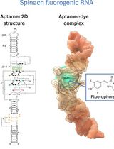
Visualizing Diverse RNA Functions in Living Cells With SpinachTM Family of Fluorogenic Aptamers
利用SpinachTM 系列荧光适配体可视化活细胞中多种RNA功能
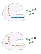
Amplification-Free Detection of Highly Structured RNA Molecules Using SCas12aV2
利用 SCas12aV2 实现高度结构化 RNA 分子的免扩增检测
神经科学
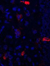
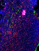
Using Combined Fluorescent In Situ Hybridization With Immunohistochemistry to Co-localize mRNA in Diverse Neuronal Cell Types
结合荧光原位杂交与免疫组织化学分析不同神经元细胞类型中的 mRNA 共定位